If hearing loss affects one ear and not the other, called unilateral hearing loss, and if the results of hearing tests indicate that sensorineural hearing loss may be causing your symptoms, doctors may recommend an MRI scan to visualize the inner ear and surrounding structures. As the tumor expands, hearing loss may worsen, facial weakness may occur, and balance problems (disequilibrium) may occur. Small tumors, which are typically limited to the bony canal, cause hearing loss in one ear, tinnitus (ringing in the ears), and unsteadiness or dizziness. What would be a potential symptom if a patient developed a tumor at the internal auditory meatus? The nervus intermedius is a branch of the facial nerve located posteriorly to the facial nerve and anterior to the superior vestibular nerve in the internal auditory canal. The facial nerve exits the internal auditory canal at the meatal foramen and continues towards the geniculate ganglion as the labyrinthine segment. Does facial nerve go through internal auditory meatus? What is a internal auditory meatus both?Īnatomical terms of bone The internal auditory meatus (also meatus acusticus internus, internal acoustic meatus, internal auditory canal, or internal acoustic canal) is a canal within the petrous part of the temporal bone of the skull between the posterior cranial fossa and the inner ear. Magnetic resonance imaging (MRI) of the internal auditory canal (IAC) is a non-invasive, painless diagnostic imaging procedure that uses using radio waves and a strong magnetic field to create detailed images of the bony canal that transmits nerves and blood vessels from the base of the brain to the inner ear. An IAM MRI scan is a useful type of MRI for investigating symptoms of earache, dizziness, tinnitus and problems with balance. These are important factors to consider when fitting earplugs.ĭue to its relative exposure to the outside world, the ear canal is susceptible to diseases and other disorders.What is a MRI internal auditory meatus both for?

On the cross-section, it is of oval shape. It has a sigmoid form and runs from behind and above downward and forward. The canal is approximately 2.5 centimetres (1 in) long and 0.7 centimetres (0.28 in) in diameter. Size and shape of the canal vary among individuals. The layer of epithelium encompassing the bony portion of the ear canal is much thinner and therefore, more sensitive in comparison to the cartilaginous portion. The bony part is much shorter in children and is only a ring ( annulus tympanicus) in the newborn.

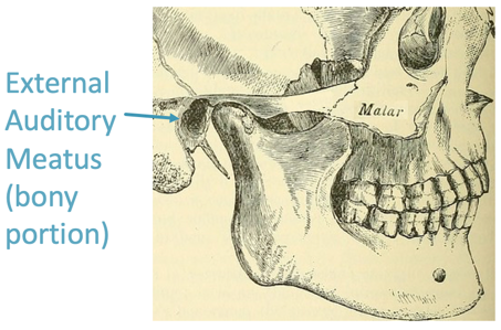
The bony part forms the inner two thirds. The cartilaginous portion of the ear canal contains small hairs and specialized sweat glands, called apocrine glands, which produce cerumen ( ear wax). The cartilage is the continuation of the cartilage framework of pinna. The elastic cartilage part forms the outer third of the canal its anterior and lower wall are cartilaginous, whereas its superior and back wall are fibrous.

The human ear canal is divided into two parts.

The adult human ear canal extends from the pinna to the eardrum and is about 2.5 centimetres (1 in) in length and 0.7 centimetres (0.3 in) in diameter. The ear canal ( external acoustic meatus, external auditory meatus, EAM) is a pathway running from the outer ear to the middle ear.


 0 kommentar(er)
